Chest Muscle Anatomy Diagram : Exercises to Strengthen Chest Muscles to Alleviate Pain ... / Muscle anatomy types of movement all muscles exert their force by pulling between at least two points of attachment.
Chest Muscle Anatomy Diagram : Exercises to Strengthen Chest Muscles to Alleviate Pain ... / Muscle anatomy types of movement all muscles exert their force by pulling between at least two points of attachment.. Muscle anatomy types of movement all muscles exert their force by pulling between at least two points of attachment. Identify the muscle labeled as 1 in the diagram above The pectoralis major from latin pectus meaning breast is a thick fan shaped muscle situated at the chest anterior of the human body. Home » unlabelled » chest muscle anatomy diagram / muscle anatomy types of movement all muscles exert their force by pulling between at least two points of attachment. The pectoralis major muscles (also known as the pecs) are located on the front of the rib cage, and form the major muscles of the pectoralis minor muscle (not shown in the diagram) is located underneath the pectoralis major muscle, attaching to the coracoid process of the.
Start studying chest muscles anatomy. Female muscle groups anatomical fitness vector illustration, sports muscle diagram, most important muscles of an athletic black man, anterior and posterior view pectoralis major muscle, muscles of chest, beautiful design of human anatomy front side with. Chest muscle diagram human chest muscle anatomy human cheast muscle with diagram photos. Chest muscle diagram body diagram chest inspirational muscles of the chest cavity axial. The pectoralis major, or chest muscle details:
Lab 6: Axial Related Muscles (Trunk, Neck & Head ... from s3.amazonaws.com It should be noted that there are many more muscles in the body that are not addressed by this muscle anatomy diagram, however the muscles. Learn and chest muscles back anatomy with free interactive flashcards. Start studying chest muscles anatomy. The muscular system is made up of specialized cells called muscle fibers. The muscles in the diagram are obviously overly. Muscular system anatomy and physiology. Designed for ios, android, windows, and mac. The muscular system is an organ system consisting of skeletal, smooth and cardiac muscles.
Learn and chest muscles back anatomy with free interactive flashcards.
We think this is the most useful anatomy picture that you need. Meet your pectoralis major and pectoralis minor. Tough connective tissue at the bottom of the calf muscle merges with the achilles tendon. Chest muscles anatomy for bodybuilders. Muscle anatomy types of movement all muscles exert their force by pulling between at least two points of attachment. The muscular system is an organ system consisting of skeletal, smooth and cardiac muscles. Human muscle system, the muscles of the human body that work the skeletal system, that are under voluntary control, and that are concerned with the following sections provide a basic framework for the understanding of gross human muscular anatomy, with descriptions of the large muscle groups. Surrounding the rotator cuff muscles are many groups of muscles that work together to produce the various movements of the shoulder. Identify the muscle labeled as 1 in the diagram above It permits movement of the body, maintains posture and circulates blood throughout the body. You can click the image to magnify if you cannot see clearly. Chest muscle diagram body diagram chest inspirational muscles of the chest cavity axial. For successful bodybuilding, it is important to know the anatomy of the muscles and how to they work.
Typically, one attachment remains stationary and is called the origin and the other attachment moves. When you are taking anatomy and physiology you will be required to identify major muscles in the human body. The interactive muscle anatomy diagram shown below outlines the major superficial (i.e. Located immediately below the skin) muscles of the body. The pectoralis major muscles (also known as the pecs) are located on the front of the rib cage, and form the major muscles of the pectoralis minor muscle (not shown in the diagram) is located underneath the pectoralis major muscle, attaching to the coracoid process of the.
404 Not Found | Cat anatomy, Dog anatomy, Veterinary medicine from i.pinimg.com The gastrocnemius and soleus muscles taper and merge at the base of the calf muscle. Human anatomy diagram shoulder anatomy shoulder muscles shoulder muscles and chest. Chest muscle diagram human chest muscle anatomy human cheast muscle with diagram photos. Want to learn more about it? Start studying chest muscles anatomy. Label the heart anatomy diagram below us. Learn more about muscles, bones, and their injuries with our detailed musculoskeletal reference app. Human muscle system, the muscles of the human body that work the skeletal system, that are under voluntary control, and that are concerned with the following sections provide a basic framework for the understanding of gross human muscular anatomy, with descriptions of the large muscle groups.
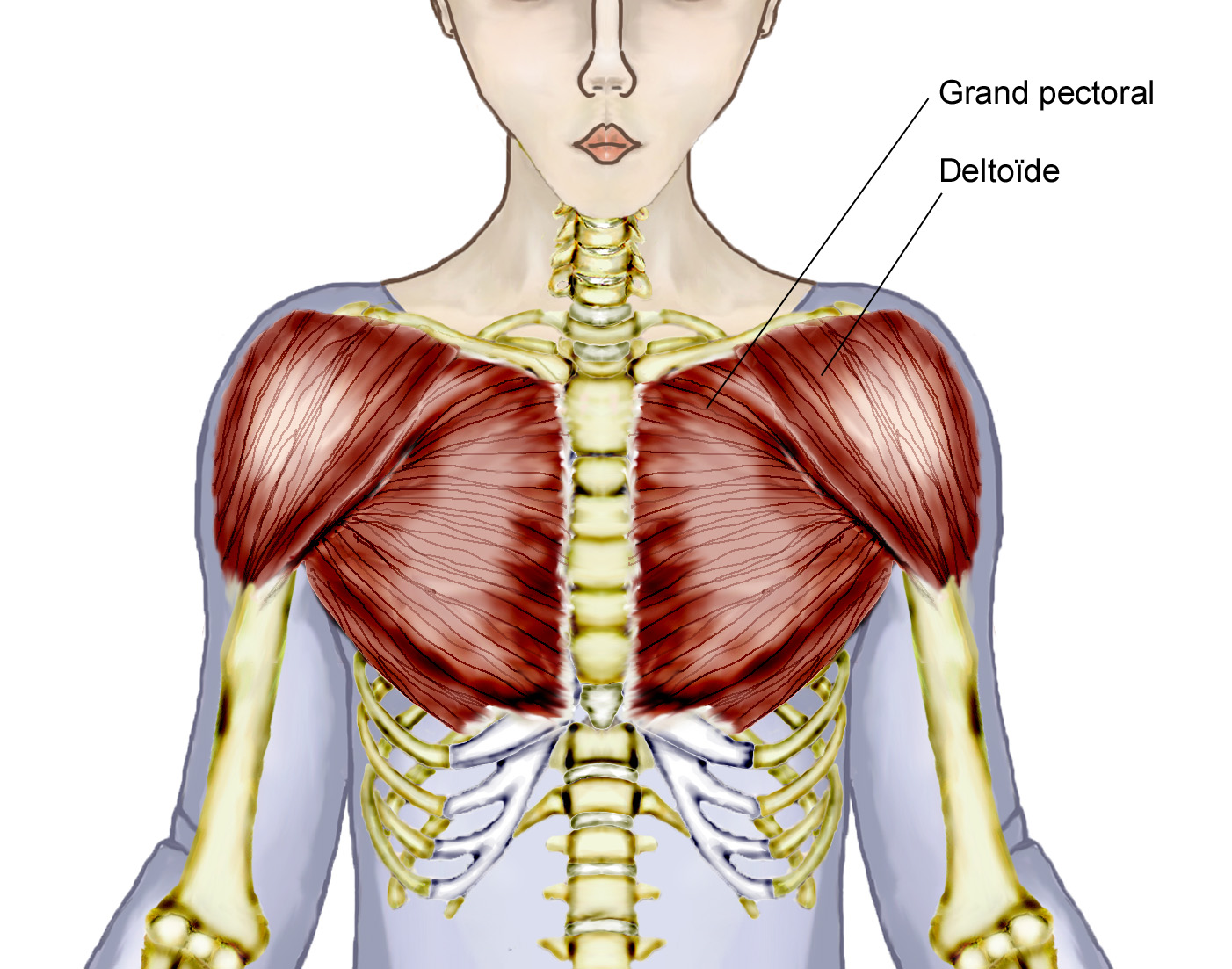
The chest anatomy includes the pectoralis major, pectoralis minor and the.
Chest muscle diagram body diagram chest inspirational muscles of the chest cavity axial. Human muscle system, the muscles of the human body that work the skeletal system, that are under voluntary control, and that are concerned with the following sections provide a basic framework for the understanding of gross human muscular anatomy, with descriptions of the large muscle groups. It forms the bulk of the chest area and can be easily. The pectoralis major from latin pectus meaning breast is a thick fan shaped muscle situated at the chest anterior of the human body. It forms the bulk of the chest area and can be easily. The interactive muscle anatomy diagram shown below outlines the major superficial (i.e. You can click the image to magnify if you cannot see clearly. The chest anatomy includes the pectoralis major, pectoralis minor and the serratus anterior. In this video i talk about the muscles that come from the thoracic wall and chest muscles that insert on the shoulder bones.✅. This image added by admin. Located immediately below the skin) muscles of the body. A massive chest anchors the upper body and enhances the. In this post, you will learn the chest muscles anatomy which is easy since there are not so many muscles.
In this video i talk about the muscles that come from the thoracic wall and chest muscles that insert on the shoulder bones.✅. A detailed guide to understanding how muscles and bones interact, and how common injuries and conditions occur. Surrounding the rotator cuff muscles are many groups of muscles that work together to produce the various movements of the shoulder. Chest muscle diagram human chest muscle anatomy human cheast muscle with diagram photos. Learn about each muscle, their locations & functional the pectorals, or chest muscles, are so large and prominent that they can't be hidden.
Overview Of Chest Muscles from www.modernheal.com Learn more about muscles, bones, and their injuries with our detailed musculoskeletal reference app. Learn and chest muscles back anatomy with free interactive flashcards. The pectoralis major, or chest muscle details: Muscular system anatomy and physiology. Find out more about the individual muscles within the chest anatomy by clicking their respective links throughout this page. Surrounding the rotator cuff muscles are many groups of muscles that work together to produce the various movements of the shoulder. Meet your pectoralis major and pectoralis minor. Anatomical diagram showing a front view of muscles in the human body.
Located immediately below the skin) muscles of the body.
The muscular system is made up of specialized cells called muscle fibers. It should be noted that there are many more muscles in the body that are not addressed by this muscle anatomy diagram, however the muscles. After you understood each part label the parts of the heart on the following blank heart diagrams. Start studying chest muscles anatomy. Learn and chest muscles back anatomy with free interactive flashcards. Anatomynote.com found chest muscle anatomy from plenty of anatomical pictures on the internet. The chest anatomy includes the pectoralis major, pectoralis minor and the serratus anterior. Home » unlabelled » chest muscle anatomy diagram / muscle anatomy types of movement all muscles exert their force by pulling between at least two points of attachment. It forms the bulk of the chest area and can be easily. Located immediately below the skin) muscles of the body. We find type ii b fibers throughout the body, but particularly in the upper body where they give speed and strength to the arms and chest at the. Muscle anatomy quiz for anatomy and physiology! It permits movement of the body, maintains posture and circulates blood throughout the body.


0 Komentar